


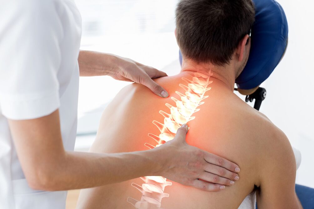
Osteochondrosis - dystrophic changes in the spine associated with age-related aging of tissues. Pathology is 80% linked to genetic data, the effect of other external factors.
Osteochondrosis- predominantly human diseases which contribute to:
- Extended service life. Over time, metabolism slows down, tissue nutrition is disrupted, and destructive regulatory systems begin to overwhelm the constructive.
- Walking straight. Standing on his feet, the person received an uneven load on different parts of the spine, he was able to perform a larger amount of movement - to twist and stretch. Abnormal lateral flexions - scoliosis - have appeared, which put an uneven load on the muscles and small joints of the spine. This increased the likelihood of developing the disease even in the class where low mobility and the rib arch protect the vertebrae - chest osteochondrosis
- Acceleration. Rapid growth makes bones, muscles and cartilage more vulnerable. The number and prevalence of blood vessels are insufficient to supply them with oxygen and essential substances
- Lack of proper physical activity. There are two extremes: sitting and car-only exercise or excessive stress in the gym when cartilage discs and cartilage wear out at an accelerated rate.
- Malnutrition. The predominance of fast carbohydrates, the lack of protein, the use of carbonated beverages lead to the body not having enough good quality building materials to maintain tissue health.
- Smoking. Causes prolonged vasospasm - disruption of tissue nutrition, acceleration of degenerative processes
- Urbanization, the large number of traumatic objects around it leads to spinal injuries, secondary osteochondrosis
Types of osteochondrosis
According to localization
- Osteochondrosis of the cervical spine
- Injury to the thoracic spine
- Lumbar osteochondrosis
- Common osteochondrosis - cervical and lumbar, thoracolumbal, lumbosacral and other combinations
In the most mobile parts, the most common lesions are the neck and lumbar. The painful spot is the transition of the moving lumbar region to the fixed sacral.
According to stage
- Initial - small changes in the middle of the disc, compaction of the nucleus, appearance of cartilage cracks
- Disease progression - cracks deepen, plate height decreases, intervertebral foramen diameter decreases. Compression of nerve roots in the spinal cord leads to pain and muscle cramps. Osteochondrosis of the spine is not only manifested by changes in the discs - due to a violation of the relative proportions of the vertebrae, the cartilage is erased unevenly on the surface of the small joints, resulting in arthrosis and arthritis.
- Complicated symptoms of osteochondrosis: further degeneration of the cartilage occurs - ruptures of the cartilage ring connecting the bodies of two adjacent vertebrae. A part of the core protrudes into the open space and compresses the roots, the spinal cord - a disc herniation is formed. A more serious problem is the separation of the lost part - a tied hernia. Severe pain, decreased sensitivity and disturbance of movement in the area for which the compressed nerve is responsible
- The body responds to increased load and excessive mobility with the growth of bone tissue - osteophytes appear. They stabilize the spine but reduce the range of motion. Bone hooks irritate muscle receptors and press on nearby blood vessels. In cervical osteochondrosis, it causes the symptoms of the "spinal artery" - dizziness, tinnitus, blinking spots in the eye
Osteochondrosis of the cervical spine
With the advent of mobile phones and computerscervical osteochondrosiseven in adolescence: a prolonged unnatural position of the head overloads the vertebrae, their discs, and joints with muscle tension.
Cervical osteochondrosis - symptoms
- Neck pain from the back of the head to the upper back
- Sometimes the headache associated with osteochondrosis of the neck mimics a migraine - one-sidedness of symptoms, intolerance to sounds and strong light, strong pulsation in the temple, bright flashes in front of the eyes
- A common headache that does not respond well to conventional pills
- Pressure drops resistant to antihypertensive drugs
- Dizziness and darkness in the eyes with sudden head turns
- Numbness of the fingers, especially after sleep, a feeling of crawling on the skin
- Restriction of movement in the neck, crackling during the movement attempt. Patients need to turn their whole bodies around to see something behind them
- Upper body sweating
- Tense muscles in the neck and shoulder girdle can be detected by touch.
If identifiedcervical osteochondrosis, treatment in the initial stage prevents serious complications - compression of the arterial vertebrae by starvation of oxygen in the brain, compression of the spinal cord.
Manifestations of osteochondrosis of the thoracic spine
Changes in the chest region are less frequent, provoking factors - back injuries, scoliosis, previous spinal diseases (tuberculosis, non-specific spondylitis, body hemangiomas).
Symptoms of a change in the thoracic region:
- Back pain - aching, pulling, worse if sitting for a long time or in an uncomfortable position. But in case of permanent pain, other possible causes must be ruled out - pneumonia, pleurisy, tumors, different types of intercostal neuralgia, herpes zoster before the bubbles appear
- Difficulty breathing, shortness of breath, inability to breathe deeply
- Chest osteochondrosis sometimes mimics an attack of angina pectoris - a person is treated for a long time by a cardiologist and the problem is in a patient’s intervertebral disc.
Lumbar and lumbosacral osteochondrosis
In the structure of all types of osteochondrosis, these classes are confident leaders, accounting for more than half of the diagnoses. This is because the greatest load falls on this area of the body, both standing and sitting. Body weight, load In case of incorrect weight lifting, in a bent position for a long time - the nucleus pulposus of the intervertebral discs is compressed, Schmorl hernias are formed through the cartilage plates. Overload and muscle cramps disrupt the position of the small joints in the vertebrae - the articular cartilage is cleared and mobility is reduced.
Several vicious circles develop at the same time: muscle cramps cause pain - the pain reflexively increases the contraction of the muscle fibers, the acute pain forces the movement to be restricted, the injured area is spared - the strength of the muscle skeleton and spinal support decreases, which increases the contraction of the muscle fibers. instability, lumbar osteochondrosis progresses.
At the mobile transition pointlumbar spineinto a single monolith of immobilized sacrum, there is a danger that the fifth lumbar vertebra will slip off the surface of the sacrum. It pinches the nerve bundles, developing radical syndrome.
Symptoms of lumbar osteochondrosis
- Low back pain, especially when sitting and standing. After resting, the horizontal position improves. In case of prolonged discharge, the pain is normal, it hurts, it pulls
- Sharp sudden lumbago when changing body condition, lifting weights, under heavy load. The patient gets stuck in the position in which he was seized, it is difficult to straighten up, to move. Lumbago is usually associated with compression of the nerve root of the spinal cord, which has developed acutely
- Transition of pain to the gluteal region, legs. The largest nerve in the body, the sciatic nerve, is a direct continuation of the spinal root, which is why patients with lumbar osteochondrosis are often worried about sciatica.
- Due to the fact that nerve fibers regulate the tone of muscles and blood vessels, regulate the nutrition of tissues, changes can be observed in the part of the trunk for which the patient’s nerve is responsible. The limb feels colder than healthy. Prolonged course of the disease can cause muscle wasting, dry skin and swelling. Local immunity is reduced - all scratches, cuts and abrasions easily become the entrance gate to infection
- Loss of sensory fibers leads to a violation of sensitivity - superficial and deep. The patient may suffer burns or frostbite because they did not experience a dangerous temperature change.
- Very frightening symptoms - numbness of the skin of the perineum, loss of control of the pelvic organs. The patient does not feel full of bladder, does not feel the need to empty the bowels. Over time, urine and feces begin to empty on their own and cannot be retained. In this case, the treatment of osteochondrosis and complications of the spine is performed surgically on an emergency basis.
Diagnosis of osteochondrosis
This is done by a neurologist or orthopedist after the therapist has ruled out pathology of the internal organs.
- The specialist finds out the main complaints, the time of their appearance, their development, the effect of drugs on the intensity of pain, rest, changes in rhythm of life
- A mandatory external examination is performed when the patient is undressing - the condition and color of the skin in symmetrical parts of the body, the tone of the tissues, the reactions to different stimuli: pain, touch, cold. or heat. Tension symptoms are indicated, indicating muscle tension and irritation of tendons and integumental membranes - fascia
- The neurological hammer reveals the uniformity and symmetry of the reflexes
- The neurologist records the volume of active (independent) and passive (performed by the doctor) movements in the joints, the ability to turn the head, the upper body without involving the lower parts of the spine.
If necessary, send it for further investigation
- Thermal imaging diagnostics
- ENMG (Electroneuromyography): Radiography. To obtain the necessary information, it must be done in at least two projections - direct and lateral. The image shows the condition of the bone tissue, the severity of the osteoporosis, the size and safety of the vertebral bodies, and reveals the osteophytes. Damaged cartilage discs are defined by the width and evenness of the intervertebral cracks. Schmorl hernia is suspected due to the unevenness of the lower or upper body. Computed tomography is recommended to clarify the nature of changes in the bone structure of the spinal column. Multispiral examination allows three-dimensional modeling of vertebrae. If necessary, MRI is prescribed to check the condition of the soft tissues - muscles, ligaments, intervertebral discs.
It should be borne in mind that the results of research should be compared with the complaints and changes observed during the investigation. Detection of signs of osteochondrosis of the spine and even disc herniation without complaints does not require more serious measures.
Treatment of osteochondrosis of the spine
Elimination of acute manifestations of the disease
- Severe pain and sharp muscle tension reinforce each other, not allowing exacerbation to subside. Therefore, the first is pain relief.
- Prescribe non-steroidal anti-inflammatory drugs by injection, muscle relaxants - muscle relaxants
- If these measures are not enough, they will block with painkillers and hormonal medications
Radio frequency denervation
Several days of bed rest are recommended
Once the symptoms have subsided, movement should be started, gradually increasing the range of motion and load. During this time, active kneading and massage are not desirable due to possible complications.
Osteochondrosis: treatment without worsening
When the patient's condition stabilizes, the normal remains sluggishosteochondrosis, the treatment consists of several components:
- Medicine. The same anti-inflammatory painkillers in tablets, capsules and ointments. A particular drug is selected by a physician based on the patient's condition, lifestyle, comorbidities, or the predominance of one or the other component of osteochondrosis. Vitamin B therapy improves the conduction of impulses along the nerve, normalizes the nutrition of tissues. Muscle relaxants continue to be used while maintaining increased muscle tone. There is no magic pill, an injection that can restore the vertebrae and cartilage to their original state. Medications relieve symptoms, improve mobility and performance. But they are unable to completely stop the progression of the disease.
- Physiotherapy. It is used to deliver drugs directly to the painful site (electrophoresis), for heating (paraffin, infrared radiation). Exposure to therapeutic currents relaxes muscles and improves nerve fiber function. After several sessions, the pain decreases and mobility is restored. It is not prescribed for active inflammation
- Manual manipulation, massage, acupuncture, acupressure. Relieve cramps by stretching and relaxing your muscles. If only the upper layer of the muscles is affected during the massage, the manual therapy penetrates deeper, so the requirements for professionals are higher. Be sure to perform an MRI first to find out the characteristics of the anatomy of that patient
- Traction. The vertebrae move away from each other, the normal distance between them is restored, the pinching of the nerves is reduced. The procedure has contraindications and should only be prescribed by a doctor
- Physiotherapy. The most effective way to treat. The only caveat is that it should be applied for life. Among the benefits - it provides activity, improves mood, increases the tone of tissues. The best methods of doctor-recommended exercises, initial yoga asanas, Pilates, swimming. They are performed smoothly, without sudden and traumatic movements, stretching the tissue, gradually increasing the amplitude
- Eating right and giving up bad habits
- Adequate supply of nutrients to tissues, good condition of blood vessels, and adequate blood supply to the vertebrae and surrounding structures prevent the progression of osteochondrosis. Proper nutrition normalizes weight, reduces spinal stress
Surgical treatment of spinal osteochondrosis.Modern clinics have a wide arsenal of minimally invasive interventions:
- Treatment and diagnostic blockade
- Radiofrequency facet ablation
- Cold plasma and laser nucleoplasty
- Endoscopic removal of a disc herniation
- Microdiscectomy
Radiofrequency thermal ablation of faceted joints
The special needles are placed exactly on the side of the intervertebral joints where the middle branch of the Lyushka nerve passes. The electrodes are built into the needles, the tip of which heats up to 80 degrees for 90 seconds. This leads to nerve coagulation. The pain goes away.
Cold plasma nucleoplasty
A special cold plasma electrode is placed on the tissue of the plate through a needle inserted into the plate. The intradiscular pressure decreases and the hernia (protrusion) extends inwards.
Microdiscectomy
In the case of a disc herniation, the adjacent nerve roots and blood vessels are compressed, with extremely painful sensations and various disturbances in the innervation of the limbs. If the effects of conservative treatment no longer prevail, for many patients, a surgical solution to a disc herniation is the only possible solution. The operation is performed under anesthesia with a 2-3 cm incision using microsurgical equipment and instruments. The duration of the surgery is 45-60 minutes. Pain syndrome is significantly reduced or completely resolved immediately after surgery in 95% of patients. The next day, the patient is allowed to walk and is soon released from the clinic.
Endoscopic removal of a disc herniation:
The hernia or free-standing sequestration is removed through the lateral intervertebral opening. A 5 mm incision is made in the skin to place the tube. The muscles, fascia and ligaments are not damaged, they are pushed apart using a tube retraction system, gradually increasing the diameter. The surgery is almost bloodless and lasts only 40-50 minutes. Patients can return to their usual treatment schedule after three weeks. The risk of complications is minimal.
Decompression and stabilization operations are performed if complications, large disc herniation, severe compression of the spinal cord nerve root, and spinal cord occur. If signs of sudden hypersensitivity, movement, or pelvic dysfunction appear, the patient should be referred to a neurosurgeon immediately. The sooner the compression can be removed, the more complete the healing will be, the person will quickly return to normal life. In this case, the surgical treatment is aimed at decompressing the compressed neural structures and stabilizing the affected segment. This is a hemi or laminectomy. The fixation is performed by a transpedicular system combined with an interstitial cage that provides 360-degree fusion. Interspinal stabilization of the vertebrae is widely used. There are many interspinous implants today. Microdiscectomy combined with interspinal stabilization, especially in the elderly, can significantly increase the effectiveness of long-term outcomes and reduce the likelihood of recurrent disc herniation.